Are Oreos Addictive? Research at Connecticut College Says Yes. And guess what even rats eat the middle first!
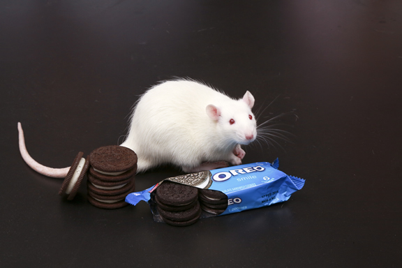
Photo by Bob MacDonnell, courtesy of Connecticut College.
Researchers at Connecticut College tested lab rats and found eating Oreos activated more neurons in the brain’s “pleasure center” than exposure to drugs of abuse.
Connecticut College students and a professor of neuroscience have found “America’s favorite cookie” is just as addictive as cocaine – at least for lab rats. And just like most humans, rats go for the middle first.
In a study designed to shed light on the potential addictiveness of high-fat/ high-sugar foods, Professor Joseph Schroeder and his students found rats formed an equally strong association between the pleasurable effects of eating Oreos and a specific environment as they did between cocaine or morphine and a specific environment. They also found that eating cookies activated more neurons in the brain’s “pleasure center” than exposure to drugs of abuse.
Schroeder, an assistant professor of neuroscience at Connecticut College, will present the research next month at the Society for Neuroscience conference in San Diego, Calif.
“Our research supports the theory that high-fat/ high-sugar foods stimulate the brain in the same way that drugs do,” Schroeder said. “It may explain why some people can’t resist these foods despite the fact that they know they are bad for them.”
Schroeder said he and his students specifically chose to feed the rats Oreos because they wanted a food that is palatable to humans and contributes to obesity in the same way cocaine is pleasurable and addictive to humans.
The research was the brainchild of neuroscience major Jamie Honohan, who graduated in May. She worked with Schroeder and several other students last year to measure the association between “drug” and environment.
On one side of a maze, they would give hungry rats Oreos and on the other, they would give them a control – in this case, rice cakes. (“Just like humans, rats don’t seem to get much pleasure out of eating them,” Schroeder said.) Then, they would give the rats the option of spending time on either side of the maze and measure how long they would spend on the side where they were typically fed Oreos.
While it may not be scientifically relevant, Honohan said it was surprising to watch the rats eat the famous cookie. “They would break it open and eat the middle first,” she said.
They compared the results of the Oreo and rice cake test with results from rats that were given an injection of cocaine or morphine, known addictive substances, on one side of the maze and a shot of saline on the other. Schroeder is licensed by the U.S. Drug Enforcement Administration to purchase and use controlled substances for research.
The research showed the rats conditioned with Oreos spent as much time on the “drug” side of the maze as the rats conditioned with cocaine or morphine.
Schroeder and his students then used immunohistochemistry to measure the expression of a protein called c-Fos, a marker of neuronal activation, in the nucleus accumbens, or the brain’s “pleasure center.”
“It basically tells us how many cells were turned on in a specific region of the brain in response to the drugs or Oreos,” said Schroeder.
They found that the Oreos activated significantly more neurons than cocaine or morphine.
“This correlated well with our behavioral results and lends support to the hypothesis that high-fat/ high-sugar foods are addictive,” said Schroeder.
And that is a problem for the general public, says Honohan.
“Even though we associate significant health hazards in taking drugs like cocaine and morphine, high-fat/ high-sugar foods may present even more of a danger because of their accessibility and affordability,” she said.